About Eye Anatomy
We think that everyone has the right to see the world accurately. That’s why we’ve chosen to focus on delivering the greatest eye care possible.
We All Deserve To See The World with Clarity.
Our eyes are one of the most precious organs we possess and a key source of our beauty. Sight is an irreplaceable gift, allowing us to perceive the world in all its vibrant colors, shapes, and sizes. Nevertheless, certain illnesses or conditions can be detrimental to our vision health leading up to blindness or sight impairment—something no person should have to experience. It’s therefore essential that we take care of this vital ability by actively protecting it from harm!
The eye serves as the bridge between our environment and our minds, transforming light into messages that are then deciphered by the brain to produce visual images. This process is made possible due to the five essential components of an eye — all measuring one inch in diameter — which reside within a cone-shaped chamber inside of a skull for protection and optimal visual development. The eyeball, a white and round structure that encompasses all four of its parts is protected by the clear dome-like cornea against dirt and external pollutants. Its coloration comes from the iris while in its center lies the pupil – an area that appears black to us.
Book Free Consultation
Book Appointment or Video Consultation online with top eye doctors
Eye Anatomy
The eye’s basic and fundamental anatomy comprises of:
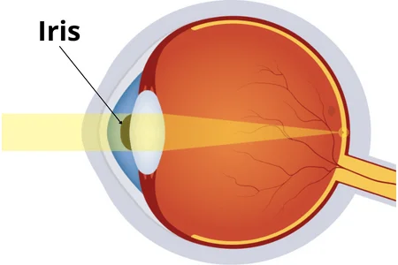
Iris
The iris – the clear front of your eye that consists of diverse hues for every individual – contains a pupil at its center, which functions as a regulator to regulate size either in contraction or dilation. This marvelous coloration is attributed to pigmentation in the stroma, an incredibly thin layer of tissue inside our eyes. The pigments can be brown, blue, green, hazel or any other hue.
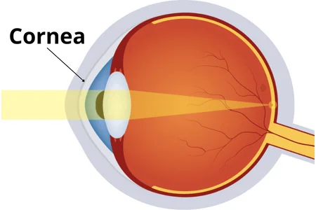
Cornea
The cornea is the protective outer layer of the eye, known for its transparency that allows light to enter and focus. It consists of five layers with a tear film providing vital nutrients and oxygen to each part. The outermost epithelium houses regenerative cells capable of swift healing from minor injuries, while the stroma forms its thickest central area – supplying strength to this key anatomical feature. The innermost layer of the cornea, endothelium—which is a single cell layer—helps maintain fluid equilibrium in the stroma. Furthermore, this part strengthens the eye and contributes to the clarity necessary for clear vision. Unfortunately, it can become blurred due to either inherited traits or outside factors such as injury or sickness.
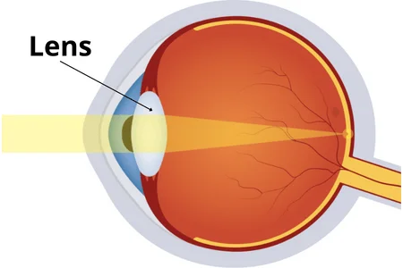
Lens
Positioned right behind the iris, our lens is a transparent structure responsible for adjusting its shape to accommodate both near and far vision. Usualy flat when at rest, it will curve inwards (convex) upon focusing on close objects thanks to ciliary muscles attached externally to the eye. These small but powerful muscles are what helps us adjust accordingly and maintain clear sight no matter how close or distant an object may be!
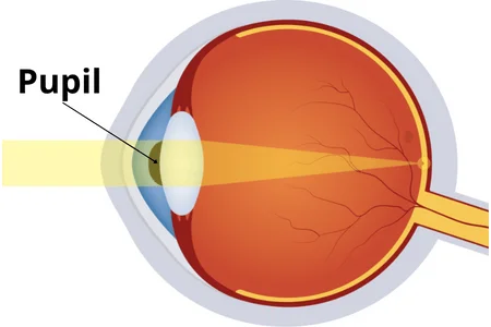
Pupil
The pupil is the blackened center of the eye, acting as a gatekeeper for how much light enters. Its hue is due to its absorbing tendencies – it takes in all visible light and reflects a red color when pictured by cameras. The pupil’s shape can be shifted depending on brightness levels; if you reduce illumination or move into darker settings, your pupils contract while they expand in brighter environments. Acting like tiny doors opening and closing with changes in lighting, these pupils protect our eyes from overexposure while simultaneously allowing us to see clearly enough no matter what scenario we’re placed in!
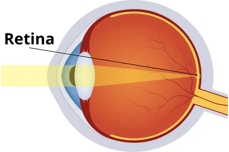
Retina
The retina, a light-sensitive tissue in the eyeball, is composed of sensitive nerve fibers and two distinct kinds of receptors that are embedded into its walls. This receptor type is color-responsive and houses rods which absorb black & white light for usage like a camera film prior to it being transmitted to your brain.